
How is lipedema diagnosed?
- Researched against current medical guidelines
- Every claim sourced & linked to a named authority
- Independent — we don’t sell surgery
- Not a substitute for your doctor.
Lipedema is a clinical diagnosis: a knowledgeable clinician identifies it from your history and a physical exam. There's no blood test or scan that confirms it. Imaging is used mainly to rule out other causes like lymphedema or vein disease.
On this page
Why is there no test for lipedema?
Lipedema is diagnosed by pattern recognition — a clinician assesses your history, the distribution and feel of your tissue, and rules out other causes. There is no biomarker, biopsy, or imaging finding that says "lipedema confirmed." This is not unusual for fat-tissue disorders and does not mean it is not real or serious.
The leading references for diagnosis are the US Standard of Care (Herbst et al., Phlebology 2021) and the Delphi Consensus (Nature Communications, 2026), which formalizes international expert agreement on diagnostic criteria.
The ~17-year gap
On average it takes about 17 years and visits to multiple doctors before a correct lipedema diagnosis is reached, according to patient survey data (Aday et al., Vascular Medicine 2024). Most delays are caused by low awareness, not by diagnostic ambiguity. Knowing the criteria — and finding a clinician who does — is the fastest way to close that gap. Don't wait.
What criteria do clinicians use?
A diagnosis of lipedema is typically made when most or all of the following are present. No single criterion is sufficient on its own — it is the overall pattern that counts.
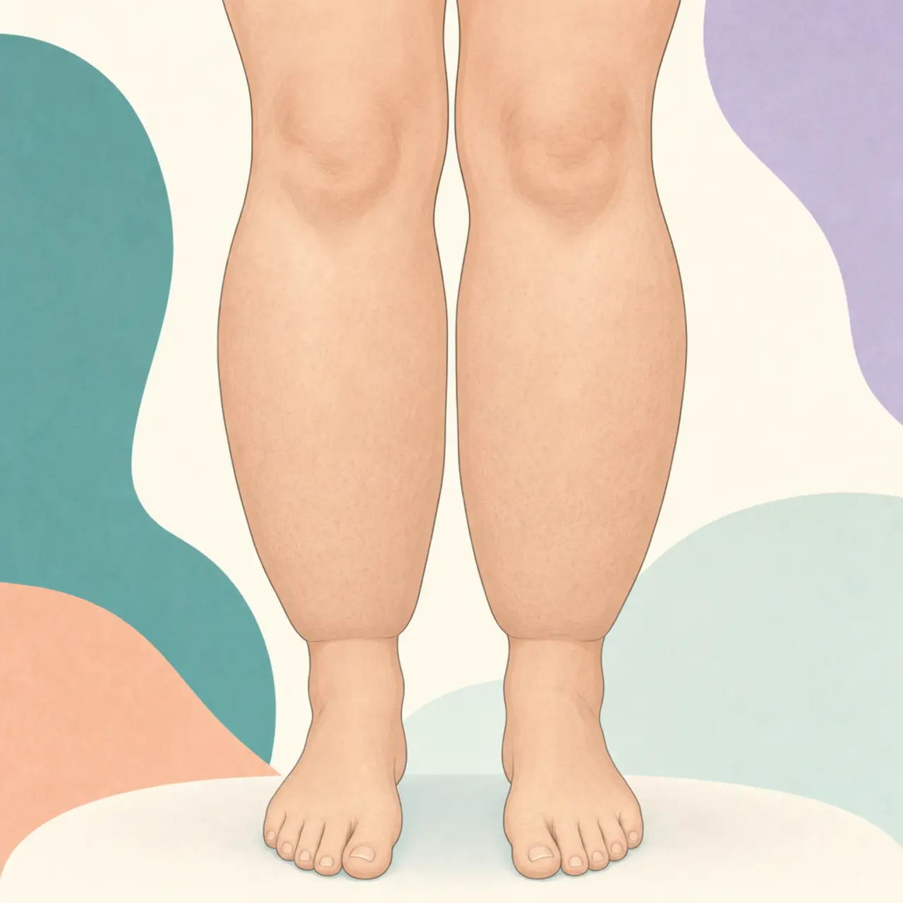
- Symmetrical, disproportionate fat in the legs (and often arms) — both sides equally affected.
- Pain or tenderness to light touch in the affected tissue (pinch pain).
- Feet and hands spared — the "cuff sign" at the ankle or wrist.
- Easy bruising — capillary fragility causing bruises from minor contact.
- Nodular texture on palpation — soft granules or nodules felt under the skin.
- Hormonal onset or worsening — started or noticeably changed at puberty, pregnancy, or menopause.
- Family history — a close relative with the same pattern.
- Diet resistance — the affected tissue does not reduce with caloric restriction.
What is the Stemmer sign, and what does it show?
The Stemmer sign is a clinical test used to detect lymphedema — not lipedema. To perform it, a clinician tries to pinch and lift the skin at the base of the second toe (or second finger):
- Positive Stemmer sign: the skin cannot be lifted — it is thickened and tethered. This suggests lymphedema is present.
- Negative Stemmer sign: the skin lifts normally. This is the expected finding in pure lipedema, because lipedema spares the feet and toes.
Critical: the Stemmer sign tests for lymphedema, not lipedema
A negative Stemmer sign is consistent with pure lipedema (feet spared). A positive Stemmer sign indicates lymphedema is also present — it does not rule lipedema out. If a clinician tells you "your Stemmer sign is negative so it's not lipedema," that is a misuse of the test. The sign tests for lymphedema; lipedema has no equivalent single physical test. Per Delphi Consensus 2026 and Standard of Care 2021.
Do I need a scan or imaging for lipedema?
Imaging is adjunct and exclusionary — it helps rule out other conditions, not confirm lipedema. Common studies ordered alongside clinical evaluation include:
- Lymphoscintigraphy or lymph MRI — to assess lymphatic function and distinguish lymphedema from lipedema when the picture is unclear.
- Venous duplex ultrasound — to rule out chronic venous insufficiency or deep vein thrombosis as a cause of leg swelling.
- Ultrasound of the soft tissue — can show the hypoechoic nodular pattern characteristic of lipedema, though this is not diagnostic on its own.
BMI is not a useful diagnostic tool for lipedema. Many people with lipedema have a normal or near-normal BMI, especially in earlier stages or with only arm involvement.
Which doctor diagnoses lipedema?
Because lipedema sits at the intersection of lymphology, vascular medicine, and dermatology, and general awareness remains low, finding the right clinician matters more than the specialty label. The following types of clinicians are most likely to be familiar with lipedema:
- Lymphologists — specialists in lymphatic disorders; most likely to be up to date.
- Vascular surgeons or phlebologists — vein and vascular specialists who often see fat and fluid swelling.
- Dermatologists with a lymphedema/fat-disorder focus.
- Knowledgeable primary care or internal medicine physicians — rare but exist; they can diagnose and refer.
- Certified lymphedema therapists (CLTs) — not physicians, so they cannot formally diagnose, but they often recognize lipedema before a physician does and can guide you to the right referral.
You may need to ask specifically
Many GPs and even some specialists have not seen enough lipedema cases to recognize it confidently. Asking for a referral to a lymphologist or vascular specialist — and bringing a printed summary of your symptoms — significantly improves your chances of a timely diagnosis.
When should I seek urgent care instead of a routine appointment?
Seek urgent care for these signs
Sudden one-sided swelling, or skin that becomes red, hot, painful, and is accompanied by fever, warrant urgent evaluation — they may indicate a blood clot (deep vein thrombosis) or a skin infection (cellulitis). These are not typical lipedema presentations and need same-day medical attention.
What should I do to get diagnosed?
The best starting point is to document your symptoms — when they started, what makes them worse, your family history, how the fat responds to diet — and bring that to a clinician who knows lipedema. Our symptom checker helps you organize this.
Sources
- Herbst KL et al. — US Standard of Care, Phlebology 2021 journals.sagepub.com
- Delphi Consensus on Lipedema — Nature Communications 2026 nature.com
- Aday et al. — US Patient Survey, Vascular Medicine 2024 pmc.ncbi.nlm.nih.gov